
What Is Magnetic Resonance Imaging?
Magnetic Resonance Imaging is a medical imaging method that uses a strong magnetic field and radio waves to create detailed pictures inside the body. An MRI can show soft tissues, nerves, joints, discs, and the spinal cord. Doctors use these images to assess possible causes of pain and guide further care.
Quick Facts About Magnetic Resonance Imaging
Category
Diagnostic imaging
Used for
Soft tissue, nerve, joint, and spine assessment
Common confusion
MRI differs from X-ray and computed tomography
Also called
MRI, MRI Scan
Often discussed with
Imaging Referrals
Key Takeaways About Magnetic Resonance Imaging
- MRI gives strong detail of soft tissues and nerves.
- Images can help connect symptoms with physical findings.
- MRI is not the same as an X-ray or CT scan.
How Magnetic Resonance Imaging Works
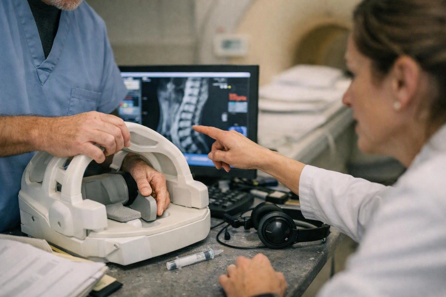
Magnetic Resonance Imaging uses a magnetic field and radio waves to produce detailed body images. The scanner detects signals from hydrogen in body tissues. A computer turns those signals into images that show different tissue types.
Related glossary terms: Computed Tomography, Disc Bulge, Nerve Root.
An MRI may show a herniated disc, swelling, a nerve problem, or changes in a joint. The images appear in slices, so a radiologist can review the area from several directions. A doctor then considers the report alongside your symptoms, examination, and medical history.
Some MRI exams use contrast material to make certain tissues easier to see. The body area being examined determines the scanner setup and the images produced.
From the Premier Injury Specialists team
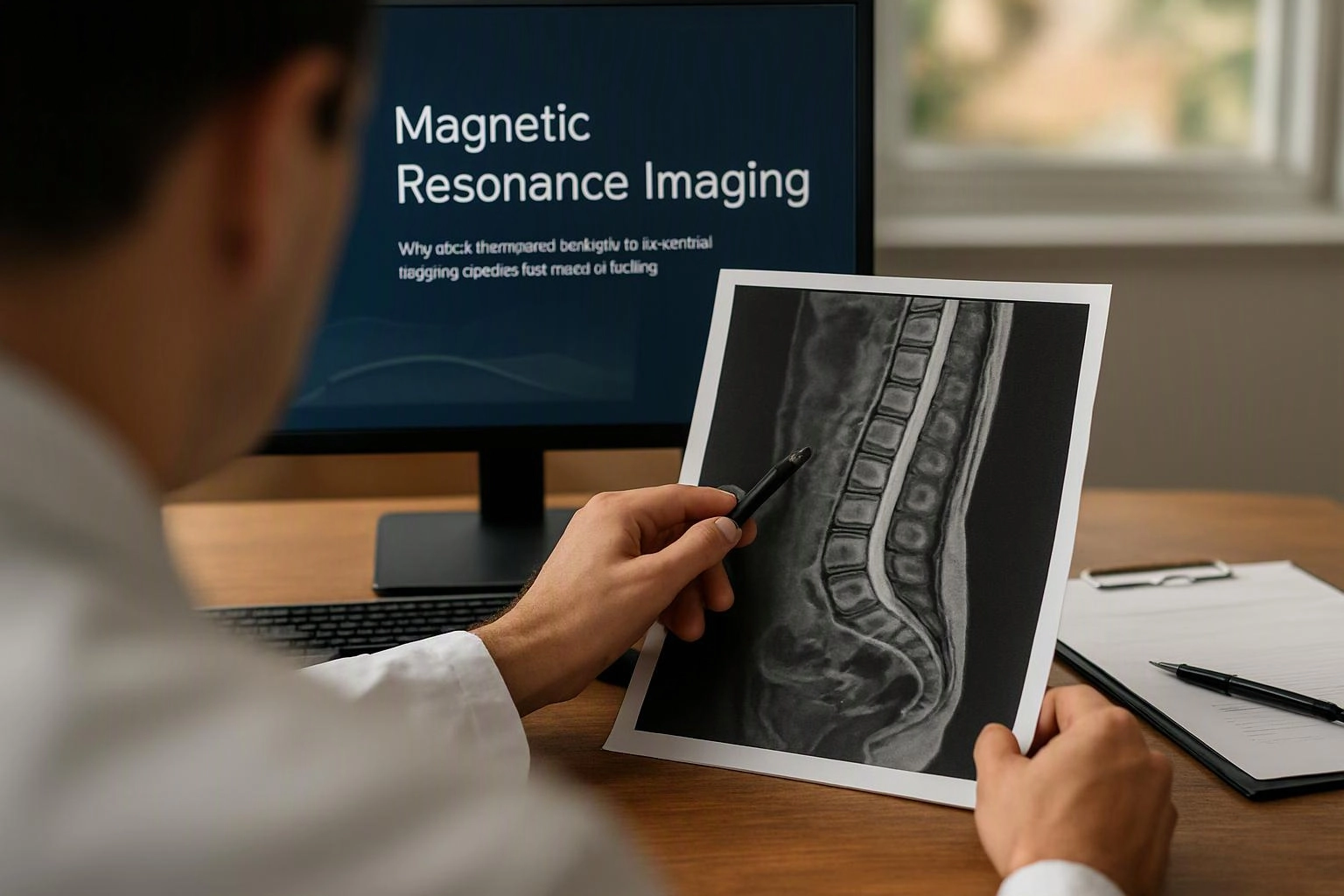
When we review an MRI referral, we compare the reported findings with the pain pattern and examination. We also check that the study addresses the body area linked to the current symptoms. Imaging can support clinical decisions, but the images do not replace a full evaluation.
Magnetic Resonance Imaging in Practice: A Real-World Example
A person with leg pain and numbness may receive an MRI referral for the lower back. The images can help show whether a disc or narrowed space may affect a spinal nerve.
Related Services
Related Terms
Computed Tomography
Computed Tomography is an imaging method that uses x-rays and computer processing to create detailed cross-sectional…
Disc Bulge
A Disc Bulge is a spinal disc change in which the disc extends beyond its usual…
Nerve Root
A Nerve Root is the part of a spinal nerve that leaves the spinal cord through…
Spinal Cord
The spinal cord is a long bundle of nerve tissue that connects the brain with nerves…
Premier Injury Specialists
Have Questions About Magnetic Resonance Imaging?
Contact Premier Injury Specialists for practical guidance on Magnetic Resonance Imaging and related pain management physician work in Stockbridge.